
AI enhanced image analysis
Boost your analysis with AI
We are bringing a whole new level of flexibility and power to your current image analysis pipeline by allowing you to embed deep learning into the process.
In Aivia 9.5, each Aivia recipe now includes an optional deep learning pre-processing step. The result of this step is then directly passed along to the recipe for further analysis, yielding a seamless way to enhance and analyze your images.
-
Enhance any Aivia recipe with a deep learning (DL) pre-processing step
-
Apply DL models as a recipe
-
Easily batch apply DL models
-
Online library of pre-trained DL models
Additionally, deep learning models can be applied to any image via the recipe console. Open or drag-and-drop the model into the recipe console and it automatically converts to a recipe for you. This provides you an easy way to deploy and batch apply any pre-trained deep learning model from our library or created in Aivia.
Using this new system you can add the power of AI smoothly into your existing analysis workflow.
Media Gallery
Image credit: Chen et al, 2020

AI image restoration
and virtual staining
Leverage our expertise
Over the last 3 years we have worked closely with top researchers to develop impactful AI models for the microscopy community. Now we are releasing these ready-to-use models to the wider community.
We are providing 20 new pre-trained deep learning models covering 7 different organelles and 3 imaging modalities. The application of these models fall into multiple categories: image restoration, super resolution, deconvolution, image segmentation and virtual staining. Most of these models are validated and characterize in our recent pre-print, check it out here.
-
20 new pre-trained DL models ready to use
-
Covering 7 organelles and 3 imaging modalities (iSIM, confocal, STED)
-
Applications include image restoration / deconvolution, image segmentation and virtual staining
-
Model library to explore and search for the right model for you
These models are available to you (the Aivia user) and can be accessed and used in multiple ways. If your data looks similar to our training data you can deploy it in Aivia as a recipe, as a pre-processing step in an application specific recipe (e.g. 3D Cell Analysis), or in Aivia Cloud. If your data is different, you can modify our model slightly (transfer learning in Aivia Cloud) or use our model structure to train a brand-new model (regular Aivia Cloud training of DL model).
You can access the pre-trained models from within Aivia (Help>Update>Models). We also organized these models (including a description and test image data) into a online library so you can explore and find the best model for your application. Just search the library for your organelle, imaging modality or application.
With this large selection of pre-trained deep learning models, you don’t have to be an expert to use AI.
Media Gallery
Image credit: Chen et al, 2020

Aivia your way
Your workspace, your set up
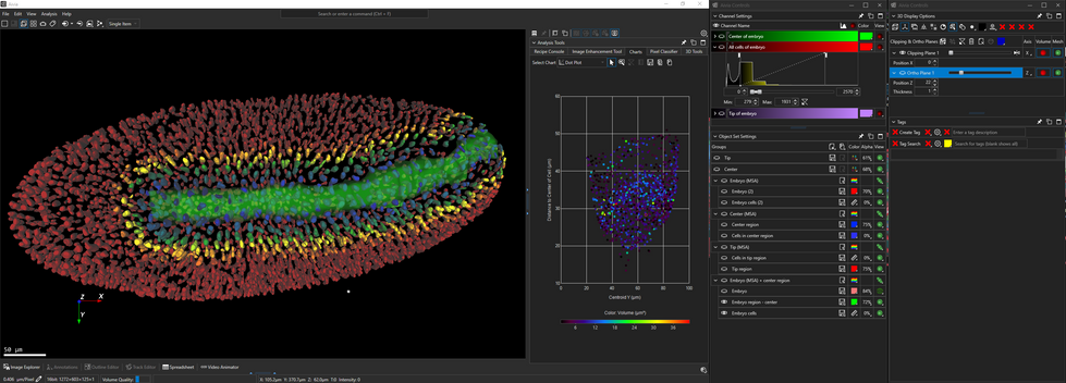
Every image analysis workflow requires a different set of tools and everyone has a preference on how to organize the tools. In Aivia 9.5, we are introducing a highly flexible GUI to help you organize everything to your taste.
All Aivia subpanels located on the right side can now be docked/undocked, pinned or place floating anywhere on 1 or more screens. Giving you the ability to arrange the tools in locations that best meets your work set-up. Multi-screen setups are also supported!
-
Ability to dock / undock, pin or float any subpanels
-
Save and reuse the subpanel location at any time
-
Multiple subpanel layouts can be saved
When you find a layout that you love, save it for future use. You can even save multiple layouts, one for each imaging analysis task such as visualization, video creation, results from manual annotations, 3D object detection, etc.
Find the right tools where and when you need them.
Media Gallery
Image credits: Neuron, pre and postsynaptic labels - Dr. Wan-Qing (University of Washington. Drosophila embryo - Dr. P. Keller (HHMI). Organoid - Dr. Ed Lachica (3i)

Tag and search
Keep your notes in context
In Aivia 9.5, we are introducing a new feature that allows you to place a text tag anywhere in your 3D/4D image. This can be used to label objects of interest, mark locations you want to revisit, or keep/share notes you have about the image.
Never lose track of your tags with our search functionality to help you quickly find the tag or group of tags you want. Additionally, the adaptable 3D view navigates to the selected tag giving you instant access to the location of each tag.
-
Add text tags anywhere in your 3D / 4D image
-
Create tag presets for project spanning multiple images
-
Search feature to quickly locate any existing tag
Tag presets makes this system ideal for annotating large, multi-image, collaborative projects. Simply create all the types of tags that you need, save them as preset and re-load them each time you need to annotate.
Label what is important to you or share your thoughts with colleagues all in the context of the image.
Media Gallery
Image credits: Neuron, pre and postsynaptic labels - Dr. Wan-Qing (University of Washington.
More features and improvements
-
New 2D Cell analysis recipe to detect cells, nuclei and up to 5 different types of organelles
-
Copy and paste selected objects to a new object set or an existing set for more ways to organize your results
-
New object visibility control - hide or only show selected objects
-
New template batch apply mode - batch apply the displayed Aivia recipe and measurement selection
-
Multiple story board per image to give you flexibility when telling your story
-
New object classifier options including training on multiple images and ability to select which object set to use in the classifier
-
Various bug fixes


