Mouse Embryo Segmentation - BioImage.io
- jose-miguelserra-l
- Jan 1, 2016
- 2 min read


Authors
A. Wolny , V. Bondarenko
Description
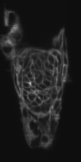
A 3D U-Net trained to predict the nuclei and their boundaries in fixed confocal images of developing mouse embryo.
First model is for fixed embryos, second model for live embryos.
Input channel: 3D Stack, grayscale
Scale: (0.2 µm × 0.2 µm × 1 µm)
Bit depth: 8-bit or 16-bit
Output channel: Model 006 - Channel 0, probability of nuclei, Channel 1: probability of nuclei contour, Model 007 - probability of cell contours
This model was downloaded and converted from BioImage.io, respecting the associated license (see License section below for more information)
Download
By downloading, installing, copying, accessing, or using the software, you agree to the terms of this end user license agreement.
Download model files and test images (ZIP archive)
Requirements
Make sure you have installed Aivia and the required DeepLearning module (according to our Wiki).
Installation and apply instructions
Aivia is required for applying the model file. You can request a demo copy of Aivia here.
Drag-and-drop the model file into the Recipe Console area; or use the 'Load recipe' option in the Recipe Console to load the model file.
Load the test image (or any image of your own) into Aivia.
If your image contains more than one channel, click on the 'Input & Output' section and specify the image channel you wish to apply the model on.
Click 'Start' to apply the model.
License
Copyright 2022, A. Wolny, V. Bondarenko. MIT License. Full license information can be found here.
Acknowledgement
Bioimage.io is supported by AI4Life. AI4Life has received funding from the European Union's Horizon Europe research and innovation program under grant agreement number 101057970.
About AI4Life: https://ai4life.eurobioimaging.eu/.
References
V. Bondarenko et al. Ex vivo Engineering Uterine Environment for Peri-implantation Mouse Development https://www.biorxiv.org/content/10.1101/2022.06.13.495767v1.full
Data: https://osf.io/uzq3w/


Comments